FAQs: MRI
MRI
What is MRI?
MRI (Magnetic Resonance Imaging) is a diagnostic imaging procedure that uses radio waves to take detailed images of human tissue. The advantage to having a MRI is that it uses radio waves, instead of X-Rays, so there is no exposure to radiation. Once the patient is ready for the MRI, a device called a coil is placed over the area to be imaged, and the procedure begins.
MRI of the brain
An MRI of the brain provides high quality two and three-dimensional images that can show both surface and deep brain structures with high clarity and accurate anatomical detail. A physician may refer you to a radiologist for an MRI of the brain when you experience:
- memory loss
- seizures
- hearing loss
- cerebral palsy
- hemorrhage
- assess tumors
- multiple sclerosis
- stroke
- and other diseases of the brain
MRI of Spine
An MRI of the spine is able to detect both normal and abnormal tissue and often makes it possible to determine the cause of back pain. An MRI of the Spine may be recommended for a patient who experiences:
- persistent back and leg pain
- to detect bulging, degenerated or herniated disks
- to pinpoint compressed, or pinched, nerves
- before surgeries to help in the planning process
- after surgery to look for changes and any scarring or infection
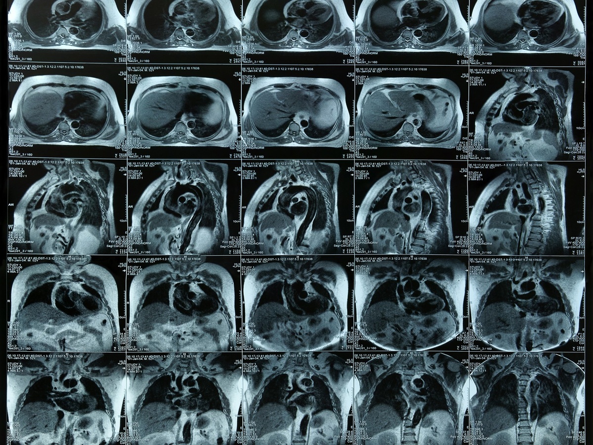
MRI of Chest, Abdomen or Pelvis
A chest, abdomen or pelvis MRI provides information on the heart and lungs, abdomen and pelvis that could not otherwise be detected from an x-ray, ultrasound or computed tomography (CT) scan.
And MRI of the chest, abdomen or pelvis may be recommended for:
- detecting abnormal growths including cancer
- determining the stages of cancer growth
- revealing problems in heart valves
- assessing problems in the ribs and sternum
What type of equipment is used for an MRI?
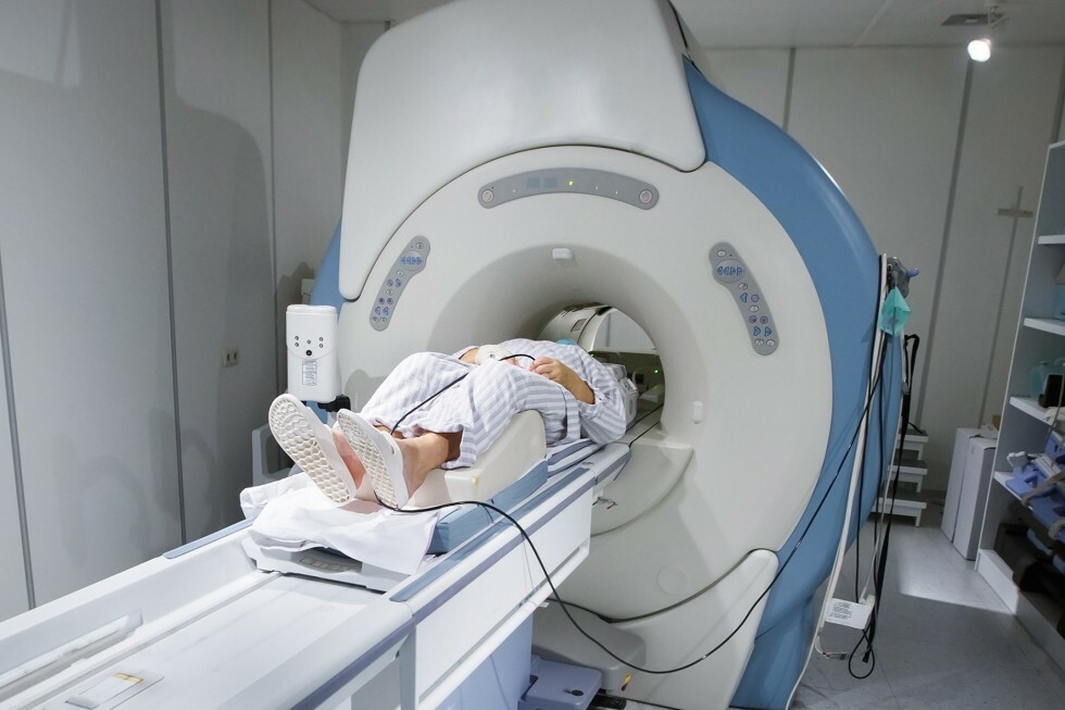
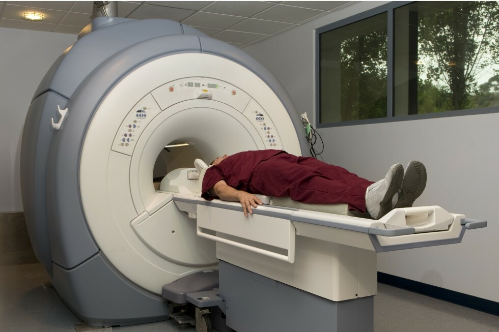
An MRI can be performed in either a closed or an open MRI scanner. A closed scanner requires you to lie in a cylinder-like compartment. An open MRI is ideal for claustrophobic, pediatric, elderly, and large patients. This MRI procedure uses a scanner that is less confining. Both types of scanners can provide your physician with accurate and detailed images of your body.
How should I prepare for my MRI?
On the day of the MRI procedure, wear comfortable clothing and try to relax. Before the MRI procedure begins, you will be asked if you have any metal medical equipment in your body such as a pacemaker, intrauterine device (IUD), implanted port, or infusion catheter. Due to the strong magnetic fields created by MRI, these devices may interrupt the MRI procedure. Also, make sure to notify the technologist if you might be pregnant. Before the MRI begins, you will also be asked to remove any metal jewelry or metal external objects as they may interfere with the procedure.
How is an MRI performed?
You will be asked to lie in the appropriate position on a cushioned table. A device called a coil will be placed on the area to be scanned. Coils are antennas used in every scan to help image the area of interest. Different coils are designed for different parts of the body and will conform to your shape as you are being imaged. You will not experience any discomfort from the coil. After you are positioned, the table will move under the magnet. The radiologic technologist will leave the room once the procedure begins to control the equipment and perform your scan. You will be able to communicate with the technologist through an intercom during the procedure. The machine will make a slight rapping sound as the images are being taken. In between scans the machine is quiet. The process takes between 30 and 45 minutes and is painless.
Will I experience any side effects from an MRI?
In some cases, your physician or the radiologist may request a contrast agent (dye) be used to improve the quality of the images. The agent is designed to make organs and blood vessels more visible, and will likely cause no side effects. You may experience a metallic taste in your mouth and in rare cases you may experience more serious side effects. The technologist can answer any questions about possible side effects.
How will I get the test results for my MRI?
The results of your MRI are read by the board-certified radiologists of Professional Radiology, Inc. A detailed report will be sent to your referring physician within 24 hours.
MRI - Brain
What is an MRI of the Brain?
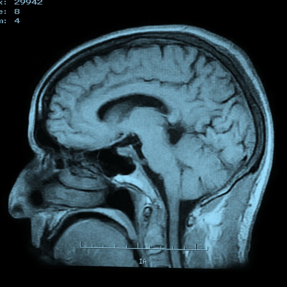
Magnetic resonance imaging, or MRI, is a method of producing detailed pictures of organs and body tissues by exposing a patient to radio waves in a strong magnetic field. The field is measured and analyzed by a computer, which forms two or three-dimensional images that may be viewed on a TV monitor. Because it uses radio waves and a magnetic field rather than x-rays, there is no exposure to radiation. An MRI of the brain provides high quality two and three-dimensional images that can show both surface and deep brain structures with high clarity and accurate anatomical detail. An MRI is used to detect changes that occur in these structures over time.
Why might my doctor recommend an MRI of the brain?
If you experience seizures, memory loss, hearing loss, or dizziness your doctor may recommend an MRI of the brain to detect, diagnose, and/or assess tumors, multiple sclerosis, stroke, dementia, hemorrhage, cerebral palsy, or meningitis in addition to other diseases of the brain. An MRI of the brain also is useful in post-operative evaluations.
MRI - Spine
What is an MRI of the spine?
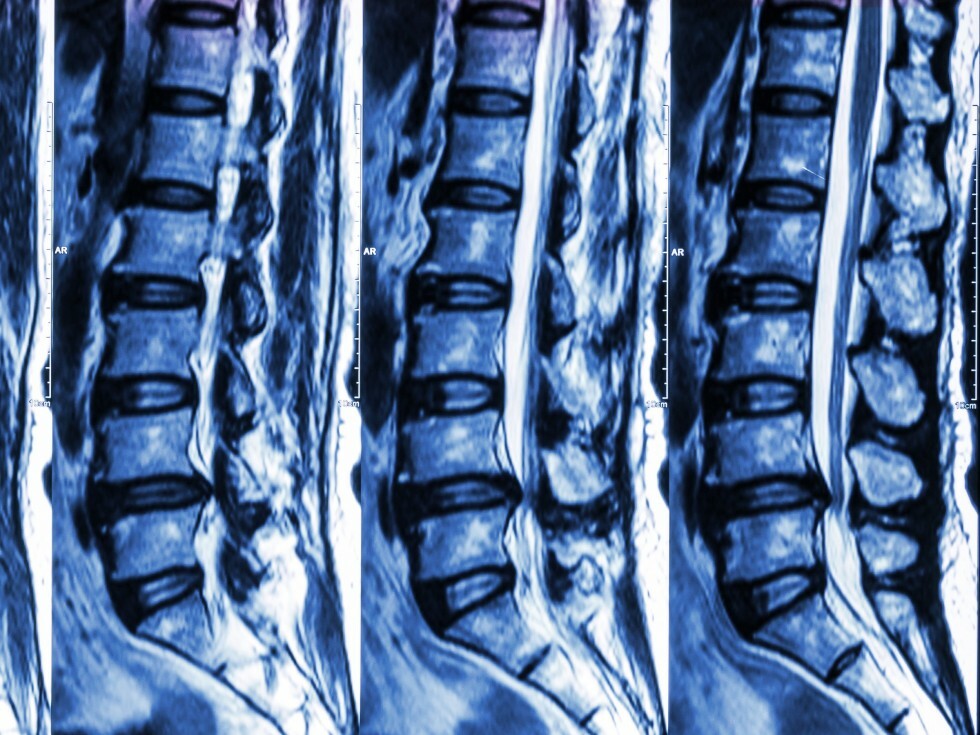
Magnetic resonance imaging, or MRI, is a method of producing detailed pictures of organs and body tissues by exposing a patient to radio waves in a strong magnetic field. The field is measured and analyzed by a computer, which forms two- or three-dimensional images that may be viewed on a monitor. Because it uses radio waves and a magnetic field rather than x-rays, there is no exposure to radiation. An MRI of the spine is able to detect both normal and abnormal tissue and often makes it possible to determine the cause of back pain.
Why might my doctor recommend an MRI of the spine?
If you experience persistent back and leg pain, an MRI of the spine may be recommended by your doctor to detect bulging, degenerated or herniated disks. It also can be used to pinpoint compressed, or pinched, nerves. Additionally, an MRI of the spine is often used before surgeries to help in the planning process and then used again after surgery to look for changes and any scarring or infection.
MRI - Chest, Abdomen
What is an MRI of the chest, abdomen or pelvis?
Magnetic resonance imaging, or MRI, is a method of producing detailed pictures of organs and body tissues by exposing a patient to radio waves in a strong magnetic field. The field is measured and analyzed by a computer, which forms two- or three-dimensional images that may be viewed on a monitor. Because it uses radio waves and a magnetic field rather than x-rays, there is no exposure to radiation. An MRI of the chest, abdomen or pelvis provides information on the heart and lungs, abdomen and pelvis that may not be detected by an x-ray, ultrasound or computed tomography (CT scan).
Why might my doctor recommend an MRI of the chest, abdomen or pelvis?
An MRI is often used to clarify previous x-rays or CT scans. Your doctor might recommend an MRI of the chest to detect abnormal growths including cancer and the stages of cancer growth. An MRI of the chest also can reveal diseased heart valves, disorders of the ribs and sternum and early breast cancer.
MRI - Joints, Extremity
What is an MRI of the joints and extremities?
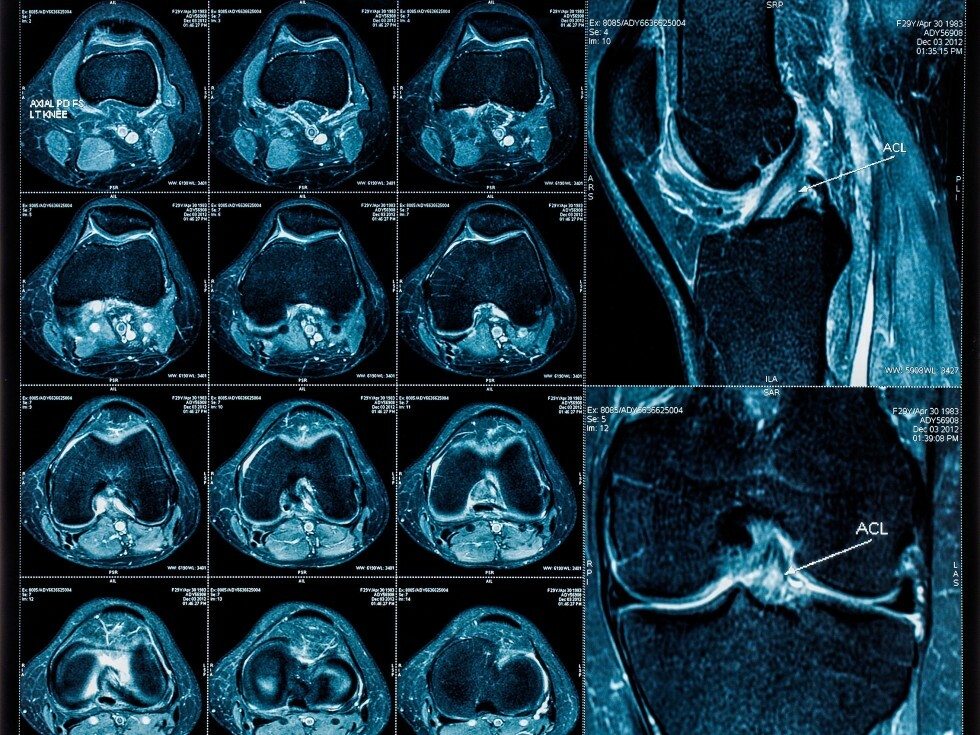
Magnetic resonance imaging, or MRI, is a method of producing detailed pictures of organs and body tissues by exposing a patient to radio waves in a strong magnetic field. The field is measured and analyzed by a computer, which forms two- or three-dimensional images that may be viewed on a monitor. Because it uses radio waves and a magnetic field rather than x-rays, there is no exposure to radiation. An MRI can be used to examine every joint in the body. Most commonly the spine, knee and shoulder are viewed, but the procedure also can be performed on the hips, wrists, and hands. An MRI is often the best choice when examining joints because it provides very clear pictures of the soft tissue near and around the bones. The procedure often is used to diagnose sports-related injuries and work-related injuries.
Why might my doctor recommend an MRI of the joints or extremities?
Your doctor may recommend an MRI of the joints or extremities to identify and locate the cause of bleeding, swelling or pain in your bones and muscles. Small tears to muscles, ligaments and tendons also can be detected. An MRI of the joints or extremities can be used to present a clear picture of degenerative disorders such as arthritis. In addition, an MRI of the joints and extremities sometimes is used to identify tumors and infections in the bone and joints and masses in the soft tissue.
MRA - Magnetic Resonance Angiography
What is an MRA?
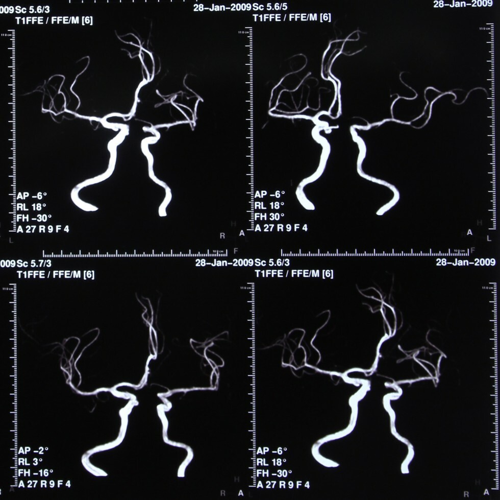
MRA stands for magnetic resonance angiography. It is an MRI study of the blood vessels. MRAs are used to assess abnormalities in the blood vessels of patients with a history of stroke, aneurysm, heart disease, and atherosclerotic vascular disease.
Why might my doctor recommend an MRA?
Your doctor might recommend an MRA if you suffer from frequent headaches. Often, MRAs also are used to evaluate problems with blood vessels such as disease and narrowing or enlargement. Additionally, MRA is helpful in detecting disease in arteries supplying blood to the brain and the development of atherosclerosis, a condition that can lead to stroke. MRA is a useful tool to detect aneurysms and evaluate your veins.
MRV - Magnetic Resonance Venography
What is an MRV?
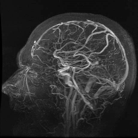
Magnetic resonance imaging, or MRI, is a method of producing detailed pictures of organs and body tissues by exposing a patient to radio waves in a strong magnetic field. The field is measured and analyzed by a computer, which forms two- or three-dimensional images that may be viewed on a monitor. Because it uses radio waves and a magnetic field rather than x-rays, there is no exposure to radiation.
MRV stands for magnetic resonance venography. It is an MRI studies of the blood vessels. MRVs are used to assess abnormalities in the blood vessels of patients with a history of stroke, aneurysm, heart disease, and atherosclerotic vascular disease.
Why might my doctor recommend an MRV?
If you experience frequent headaches, an MRV of the head may be recommended to detect or rule out blood clots in the brain. An MRV of the chest is used to detect blood clots or blockages in the main arteries leading to the heart, and an MRV of the abdomen checks for blood clots or blockages in the liver.